
Figure 3. Experimental results and mathematical model predictions of EGFR clustering.
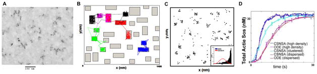
(A). Experimental evidence for EGFR clustering in absence of ligand. Electron micrograph of gold particle-labeled EGF receptors in resting A341 cells (~2 million EGFR/cell), reveals a non-random distribution and provides evidence for receptor co-confinement. (B). Spatial domain used in lattice-free Monte Carlo simulation.[28] The spatial domain simulated by the off-lattice Monte Carlo procedure was a square of area 2 μm2, representative of a small region in the plasma membrane. This region was modified to include many islands or preferred domains (the green rectangles within the membrane patch), to simulate the receptor-trapping micordomains seen in (A). Movement of receptors into and out of the simulated microdomains over a time period of 30 s is indicated by the thin colored tracings. Receptor trapping in the microdomains was reproduced mathematically by stipulating that receptors had a greater probability of entering these regions than of leaving them. (C) Simulation predictions of receptor clustering in absence of ligand. The predicted particle positions after 30 s of simulation time are indicated by the black dots. The Hopkins statistical test (inset) was used to test the randomness of receptor distribution. The right shift of the distribution (compared to the random or normal distribution shown in red) towards unity confirms the clustered nature of the receptors. The predicted receptor distribution compares well with the experimental observation in (A). (D) Simulations using a Coupled Spatial/Nonspatial Stochastic Algorithm (CSNSA) support the conclusion that EGFR clustering promotes activation of the adaptor SOS. ODE models confirm this conclusion, using a fast diffusion coefficient to override contributions from membrane spatial organization. (From Hsieh et al.[28] and Costa et al.24)