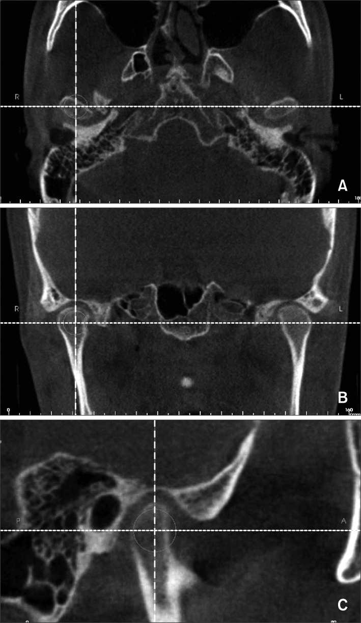
Figure 1.
The 3 views of the condyle in the cone-beam computed tomography (CBCT) image: A, axial view; B, coronal view; C, sagittal view. The CBCT images were reoriented with the horizontal reference plane connecting the bilateral orbitales and Frankfurt horizontal plane,17,18 and the vertical midline and horizontal reference planes were set accordingly. The sagittal slice (C) was evaluated at the point where the mediolateral diameter of the right or left condyles was greatest (A) in the axial view.

