Figure 4.

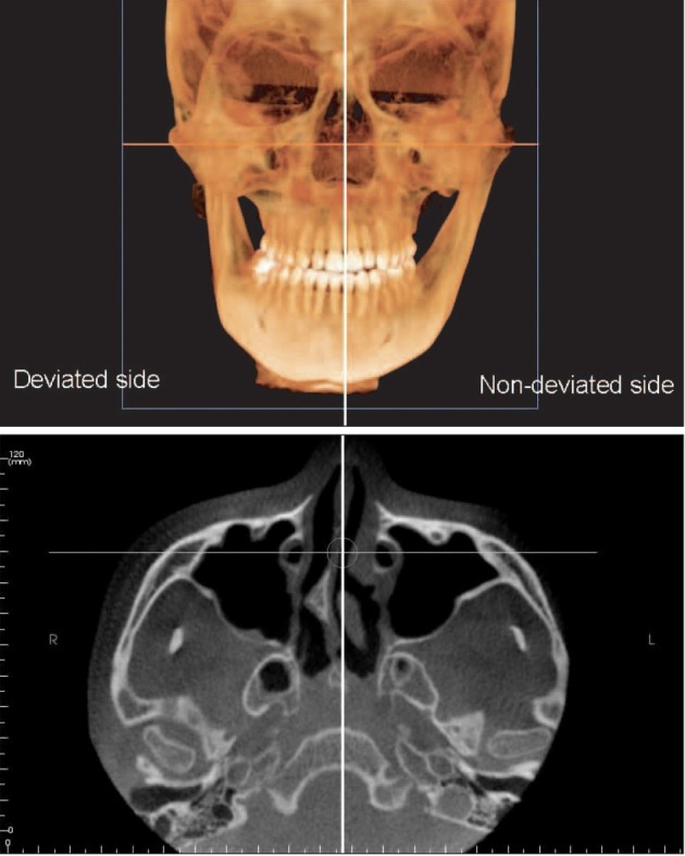
Cone-beam computed tomography image of a sample case showing the largest axial condylar angle difference between the deviated and non-deviated sides.

Cone-beam computed tomography image of a sample case showing the largest axial condylar angle difference between the deviated and non-deviated sides.