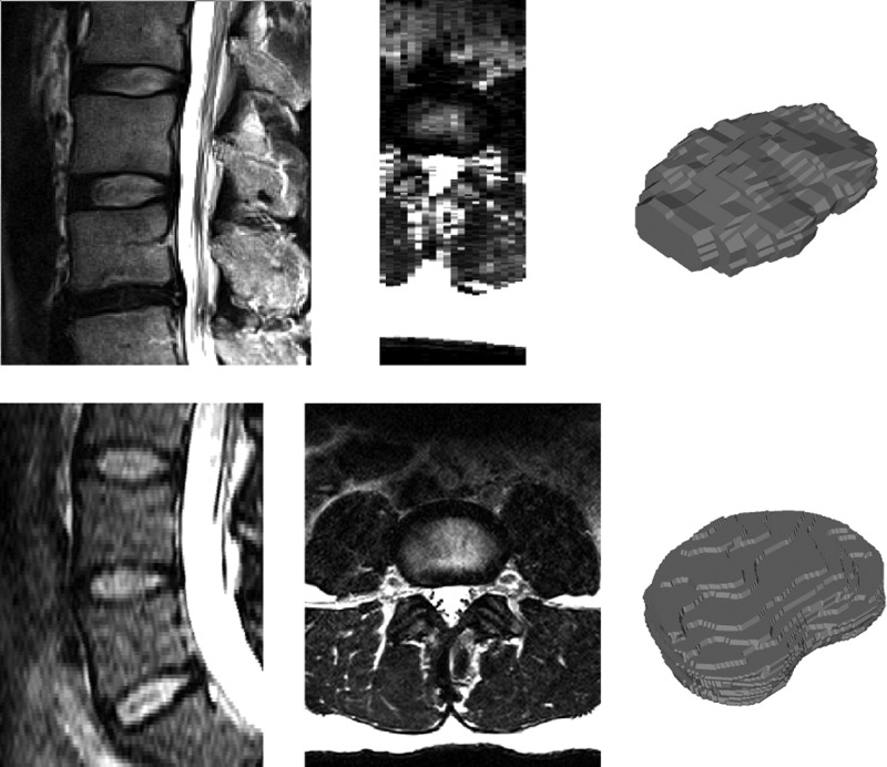
Figure 1.
Sagittal (left) and axial (middle) views of an example 2D turbo spin echo (top, 3.3 mm slice spacing) and 3D SPACE (bottom, 1 mm slice spacing) MRI scans with manually segmented IVD shapes (right). The high resolution 3D image provides information about the anatomical shape that is not available in the sparser 2D scan. IVD, intervertebral disc; 2D, two dimensional; 3D, three dimensional.

