Figure 2.

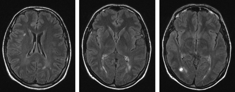
Axial fluid attenuated inversion recovery images of the identified radiologically isolated syndrome patient illustrating the multiple T2 hyperintensities and the contrast-enhancing lesion in the far right image.

Axial fluid attenuated inversion recovery images of the identified radiologically isolated syndrome patient illustrating the multiple T2 hyperintensities and the contrast-enhancing lesion in the far right image.