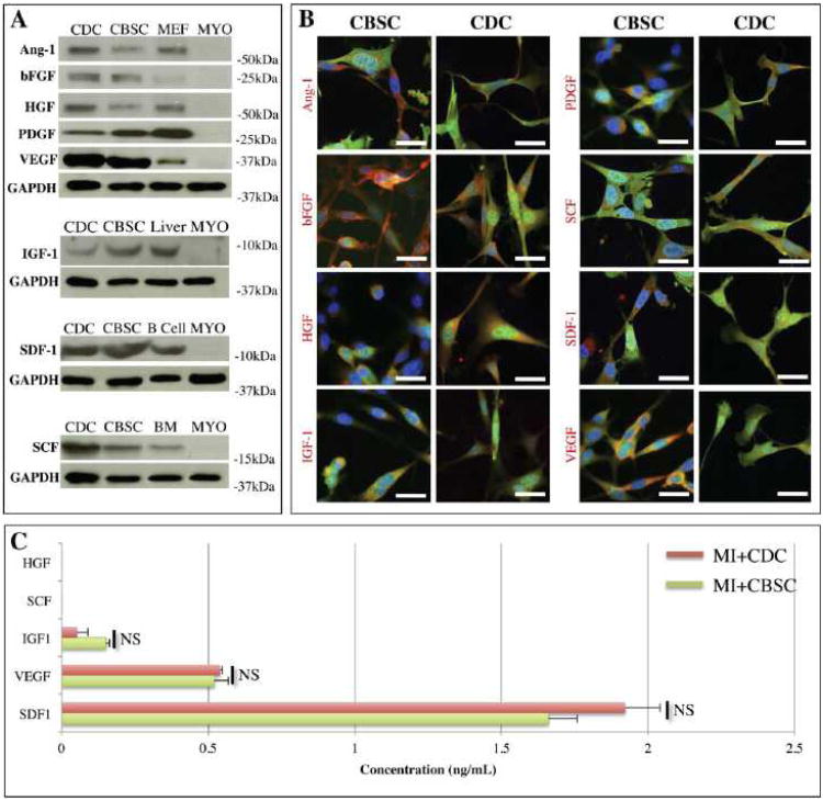
Figure 1. In vitro characterization of stem cells.
A) CBSC or CDC lysates were analyzed by Western analysis. Positive controls include mouse endothelial fibroblasts (MEF), liver, bone marrow (BM), and B lymphocytes (B Cell). Myocyte (MYO) lysates were used as negative controls for all samples. B) CBSCs (green) were fixed in vitro and immunostained against each paracrine factor (red). Nuclei are labeled with DAPI (blue) and scale bars = 20 μm. C) CBSCs or CDCs were allowed to proliferate over 72 hours and their culture media was analyzed by ELISA for the presence of soluble HGF, IGF, SCF, SDF-1, and VEGF. Samples were analyzed in triplicate and background signal was subtracted using unconditioned media blanks. NS = No Significant difference (p > 0.05).

