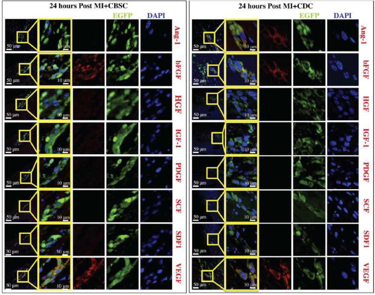
Figure 5. Characterization of paracrine factors secreted by stem cells after 24 hours post-MI in vivo.
Animals receiving MI+CBSCs or MI+CDCs were sacrificed 24 hours post-MI for analysis of paracrine factor production. EGFP+ CBSCs stained positive for bFGF, and VEGF (shown in red), but negative for HGF, IGF-1, PDGF, SCF, and SDF-1. EGFP+ CDCs stained positive for Ang-1 in addition to bFGF and VEGF, but also stained negative for HGF, IGF-1, PDGF, SCF, and SDF-1. Nuclei are labeled with DAPI (blue) and injected CBSCs are green.

