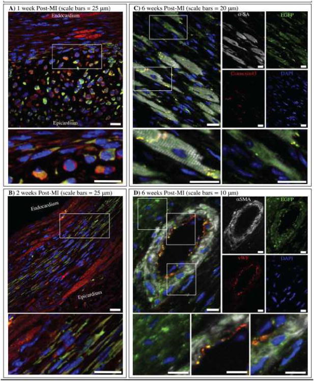
Figure 7. Cortical bone stem cells grow and differentiate over 6 weeks.
A) 1 week post-MI and B) 2 weeks post-MI samples were immunostained for α-sarcomeric actin (red) and EGFP (green). 6 weeks post-MI samples were immunostained for C) α-sarcomeric actin (white), connexin43 (red) and EGFP (green) or D) α-smooth muscle actin (white), von Willebrand Factor (red) and EGFP (green). Nuclei were labeled with DAPI (blue) in all images.

