Fig 5.

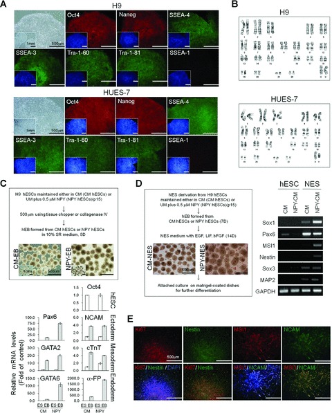
Long-term, feeder-free hESC culture in NPY medium. (A) Morphology of a representative hESC colony and immunohistochemical analysis of OCT4, NANOG, SSEA-4, SSEA-3, TRA-1–60 and TRA-1–80. H9 and HES-7 cells were cultured in NPY medium for 15 passages. Nuclei were visualized by DAPI staining (insets). Bar = 500 mm or 1 mm (inset images). (B) Karyotype analysis of hESCs cultured in NPY medium for 15 passages. (C) hEB formation and differentiation. hEBs were derived from H9 cells cultured in CM or NPY medium for 15 passages. Upper panel; representative images of hEBs. Bar = 500 mm or 1 mm (inset images). Lower panel; real-time RT-PCR analysis for the expression of OCT4 and differentiation markers characteristic of the three germ layers; ectoderm (PAX6 and NCAM), mesoderm (GATA2 and cTnT) and endoderm (α-FP and GATA6). The data are presented as the mean ± S.E. of three independent experiments. (D) NESs derived from hESCs. Left panel; representative images of NESs. Right panel; RT-PCR analysis of neural stem cell marker expression in NESs. (E) Representative images of NESs stained either for Ki67 (cell proliferation marker), nestin, MSI1 and/or NCAM. Bar = 500 mm.
