6.

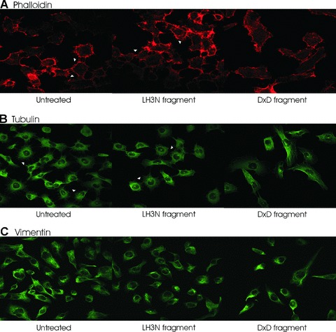
Cytoskeleton protein staining of HT-1080 cells after treatment of the LH3 N-terminal fragment with (LH3N fragment) or without (DXD fragment) glycosyltransferase activities. (A) Filamentous actin was visualized by Alexa Fluor 568 phalloidin (red). The filopodia indicated by arrowheads were well-developed in the control and the LH3N fragment treated cells, whereas the filopodia protrusions at the cell periphery were severely disrupted in the DXD fragment treated cells. (B) The overall architecture of the microtubule network was not considerably altered. However, brighter perinuclear tubulin staining around the centrosome, with much less staining extending towards the filopodia, was observed in the DXD-treated cells than in the untreated controls and in the LH3N fragment treated cells (indicated by arrowheads). (C) No obvious difference of vimentin staining was seen in the treated cells compared to the controls.
