5.

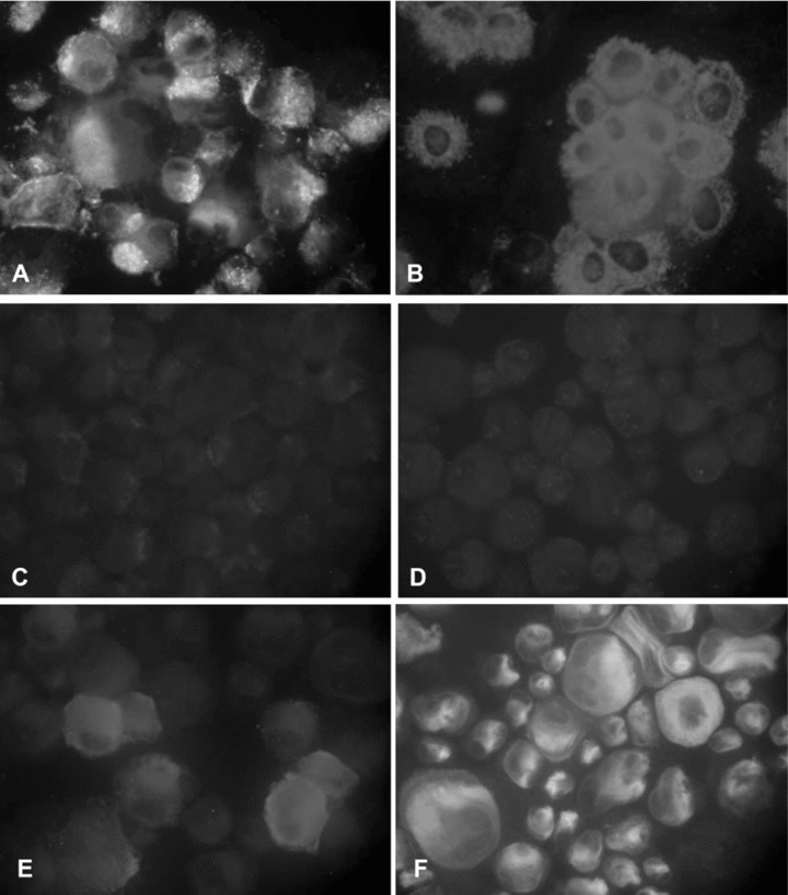
Immunofluorescence analysis of EPC-derived cells. Cells were positively stained for the panendothelial marker CD31 (A) and vWF (B), but negatively stained for the lymphatic cell-specific markers podoplanin and Prox-1 (C, D). Note that some few EPC-derived cells showed expression of the lymphatic cell-specific marker LYVE-1 (E, F). Original magnifications: (A, B, E, F) ×400, (B, C) ×200.
