4.

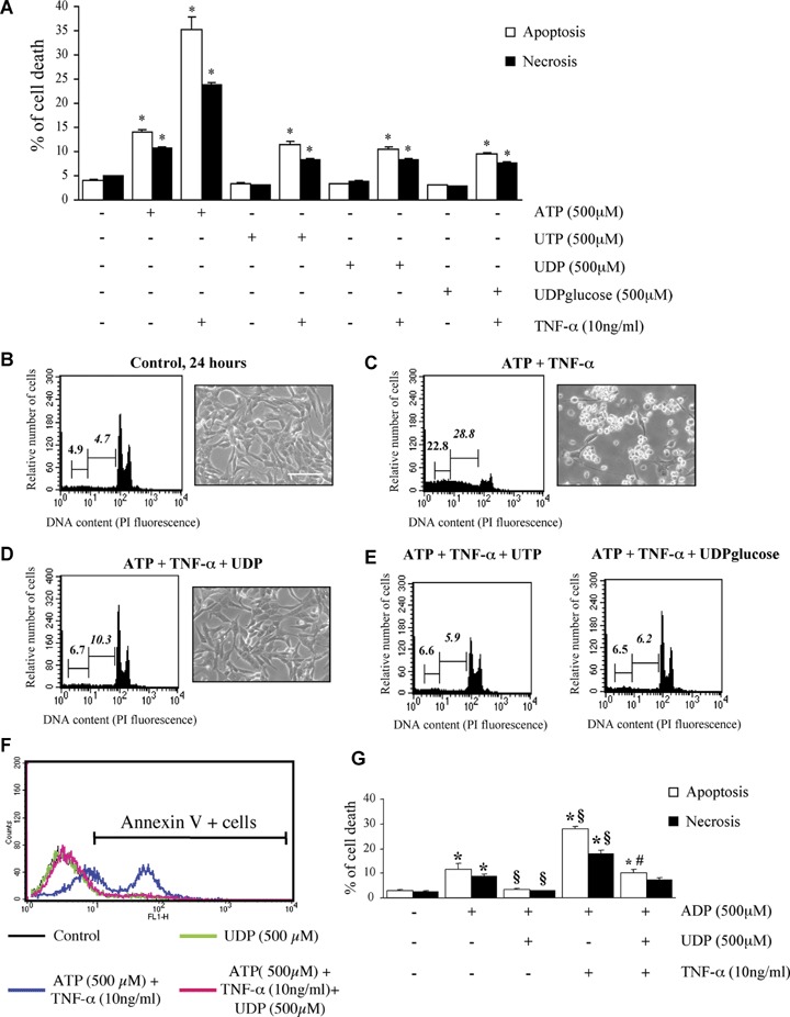
Uracil nucleotides have no effect ‘per se’ on cardiomyocytic viability, but protect cells from ATP and ADP-induced cytotoxicity. To assess the effect of uracil nucleotides on HL-1 cardiomyocytic viability, cells were exposed to UTP, UDP or UDPglucose (500 μM) alone or in combination with TNF-α, and apoptosis and necrosis determined after 24 hrs by flow-cytometry. Parallel cultures were treated with ATP, here utilized as a positive control (A). The effect of TNF-α alone is shown in Figure 2B.*P <0.05 versus corresponding Control, by one-way ANOVA (Scheffe's F test). To assess the ability of uracil nucleotides to counteract cardiomyocyte death induced by ATP+TNF-α, cells were treated with either vehicle (B), or ATP+TNF-α (500 μM and 10 ng/ml, respectively) in the absence (C) or presence of UDP (100 μM, D), UTP (25 μM) or UDPglucose (10 μM) (E). In these experiments, uracil nucleotides were added to cells 30 min before adenine nucleotides. After 24 hrs, micrographs of adhering cells were taken (scale bar: 50 μm) and flow cytometric analysis was performed on the total cell population. In cytograms, first and second numbers on the left represent the percentage of necrotic and apoptotic cells, respectively. Results from a typical experiment are shown in (B–E). Similar data were obtained in five independent experiments. UDP similarly protected cells from the apoptosis and necrosis induced by either ADP alone (500 μM) or ADP and TNF-α (10 ng/ml) (F). Annexin V staining of cells confirmed the complete protection exerted by uracil nucleotides. The percentages of Annexin V-positive cells in this representative experiment (out of three) were: Control, 8.1%; UDP alone, 8.4%; ATP+TNF-α, 56.4%; ATP+TNF-α+UDP, 11.3%. (G) *P <0.05 versus corresponding Control, §P<0.05 versus ADP alone, #P <0.05 versus ADP+TNF-α, by one-way ANOVA (Scheffe's F test); the mean values ± S.E.M.of four independent experiments are shown.
