
6.
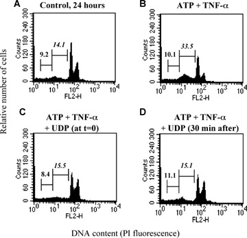
Characterization of UDP-induced protection. HL-1 cardiomyocytes were exposed to either vehicle (Control, A), ATP (100 μM) and TNF-α (10 ng/ml) (B), as indicated, for 24 hrs. UDP (100 μM) was added to cultures either together with ATP (C) or 30 min after its addition (D). At the end of treatment, flow cytometric analysis was performed on the total cell population. In cytograms, first and second numbers on the left represent the percentage of necrotic and apoptotic cells, respectively.