
5.
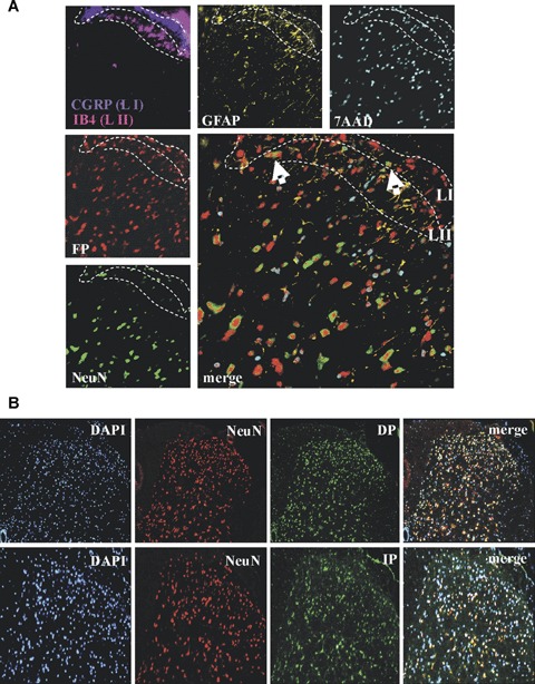
Prostaglandin receptors DP, FP and IP are expressed in dorsal horn neurons.(A) Immunohistochemical analysis of a L4–L5 spinal cord section from wild-type mice using the MELC technology. Signals are shown in false colors. CGRP (blue) was used as marker for lamina 1 (LI) and IB4 (pink) as marker for lamina 2 (LII). Arrows indicate FP-expressing neurons (white), or FP-expressing non-neuronal cells (yellow).(B) Immunohistochemical analysis of PGD2 (DP) and prostacyclin (IP) receptors in the spinal cord. Sections are from spinal cords (L4–5) of zymosan-treated (8 hrs) wild-type mice. DAPI was used for nuclear staining (blue), NeuN to stain neurons (red) and specific antibodies for the indicated prostaglandin receptors (green). The ipsilateral dorsal horn is shown.