2.

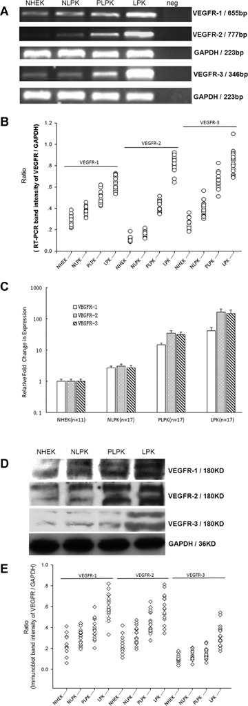
mRNA and protein levels of VEGFRs determined by RT-PCR, qPCR and Western blot, respectively, in cultured normal and psoriatic epidermal keratinocytes. (A) VEGFR-1, band at 655bp; VEGFR-2, band at 777bp; VEGFR-3, band at 346bp; GAPDH at 223bp served as an internal control for mRNA. (B) Scatter plots for RT-PCR band intensity ratio of targeted VEGFR gene versus GAPDH using Gel-Pro Analyzer software V4.0 (Media Cybernetics, USA). Expression of VEGFRs mRNA is statistically significant different between LPK and other indicated cells (P <0.001). (C) Relative quantitation of VEGFRs expression levels in NHEK, NLPK, PLPK and LPK by real-time RT-PCR. The expression of VEGFRs was normalized to the endogenous control GAPDH. The relative fold change of VEGFRs in NLPK, PLPK and LPK was compared with NHEK. The level of VEGFRs in LPK is significantly higher than that in NHEK, PLPK and LPK (P <0.001). Bars: mean±SEM. (D) The anti–VEGFR-1, anti–VEGFR-2 and anti–VEGFR-3 antibodies show bands at about 180 and 200KD. GAPDH served as a loading control for protein normalization. (E) Scatter plot analysis of protein levels of VEGFRs versus GAPDH as determined by Western blot. Expression of VEGFRs protein is statistically significant different between LPK and other indicated cells (P < 0.001). NHEK, normal human epidermal ker-atinocytes (n= 11); NLPK, non-lesional psoriatic keratinocytes (n= 17); PLPK, perilesional psoriatic keratinocytes (n= 17); LPK, lesional psoriatic keratinocytes (n= 17); neg, negative control.
