2.

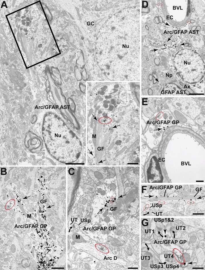
Electron micrographs from the DG showing immunoperoxidase localization of Arg3.1/Arc (red ellipses) within immunogold GFAP positive glial cell bodies (Arc/GFAP AST) and glial processes (Arc/GFAP GP; arrows). A: GFAP positive astrocytes located in the outer layers of the GC layer showing Arg3.1/Arc peroxidase reaction product in close association with the glial filaments (GF; boxed region). B and C: Arg3.1/Arc immunoperoxidase labelling within medium-sized GFAP positive glial profiles (arrows) within the ML directly associated with the plasma membrane as well as with the glial filaments (GF). In (C) we can also observed the typical dendritic presence of peroxidase-labelled Arg3.1/Arc (Arc D) which gives rise to an unlabelled spine (USp) that receives an asymmetric synapse from an unlabelled axon terminal (UT). D and E: Arg3.1/Arc Peroxidase reaction product within the cytoplasm of glial processes apposed to the basal membrane of endothelial cells (EC) lining the blood vessel lumen (BVL). Peroxidase labelling for Arg3.1/Arc appears within the cytoplasm and along discrete segments of the plasma membrane. In D the Arg3.1/Arc-labelled glial processes extending towards the BVL are proximal to the astrocytic cell body. We can also see that the Arc/GFAP astrocyte is in the vicinity of a single labelled GFAP astrocyte but separated by abundant neuropil (NP) and a myelinated axon (Ax). F and G: Peroxidase labelling for Arg3.1/Arc within the cytoplasm and along discrete segments of the plasma membrane of peri-synaptic glial profiles. The glial processes appose unlabelled dendritic spines (USp). All spines receive perforated (as in F) and unperforated asymmetric synapses (in G; curved arrows) from unlabelled terminal (UT). GC = granule cell, GP = glial profile, M = mitochondrion, Nu = nucleus. Scale bars = 2 μm in A–D, 1 μm in B–F, 0.5 μm in C–E, 0.3 μm in G and 0.8 μm in boxed region.
