1.

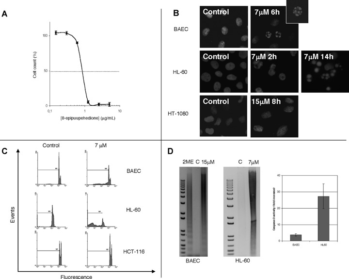
8-epipuupehedione inhibits HL-60 cell proliferation (A) and induces endothelial bovine aortic endothelial cells (BAEC) and leukaemia (HL-60), but not tumour (HT-1080 and HCT-116) cell apoptosis (B, C and D), by activation of caspase activity (D). For the study of the dose-dependent effect of 8-epipuupehedione on the in vitro growth of HL60 cells, the MTT dye reduction assay was used (A). Cell proliferation is represented as a percentage of control-cells growth. Each point represents the mean of quadruplicates; SD values were always lower than 10% of the mean values and are omitted for clarity. A typical curve is represented out of four independent experiments. For the study of nuclear morphologic changes induced by 8-epipuupehedione (B), treatments were carried out for the indicated times and Hoetsch staining was carried out as described in Material and methods section. For cell cycle analyses (C), BAEC, HL-60 and HCT-116 cells were treated with 8-epipuupehedione (7 μM) for 14 hrs, fixed with 70% ice-cold ethanol, stained with propidium iodide and submitted to flow cytometry, as described in Material and methods section. Sub-G1 area is indicated as M1. For DNA internucleosomal fragmentation and caspase-3 activation analyses (D), BAEC and HL-60 cells were treated with 8-epipuupehedione for 14 hrs. For DNA fragmentation assay, cells were harvested and centrifuged and pellets were frozen in liquid nitrogen. Internucleosomal fragmented DNA was isolated and visualized by agarose gel electrophoresis. BAE cells treated with 2-methoxiestradiol (2ME) for 24 hrs were used as positive internal control. C, control, untreated cells. Caspase-3 activity assay was carried out as described in the Material and methods section. Data are represented as fold-increases of control values. Data from duplicate samples were used in each experiment.
