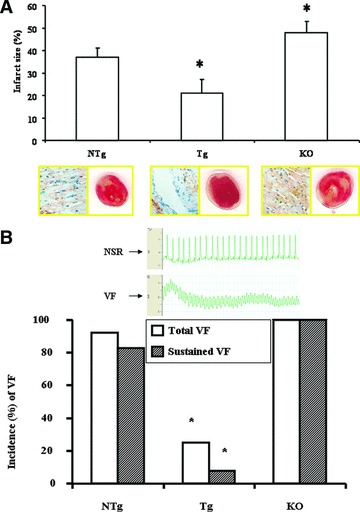
Fig 2.

Infarct size and HO-1 staining in isolated NTg, Tg and HO-1 KO mouse hearts subjected to 20 min. of normotermic global ischemia followed by 120 min. of reperfusion. Infarct is represented by white area surrounding by the living red tissues (A, right lower part). Mean ± S.E.M., *P < 0.05 compared to the NTg control values. Immunochemical localization of HO-1 is stained by blue in NTg, Tg and KO myocardium (A, left lower part). (B) Representative ECG records corresponding to sinus rhythm and VF, respectively, are given in the upper part of (B). The incidence (%) of total (open bars) and sustained (hatched bars) reperfusion-induced VF in isolated NTg, Tg and KO mouse hearts are shown in the lower part of (B). The incidence of reperfusion-induced VF was registered, and comparisons were made to the values of NTg group. n= 12 in each group, *P < 0.05. Because of the non-parametric distribution in the incidence of total and sustained VF, the chi-square non-parametric test was used to compare individual groups.
