
Fig 1.
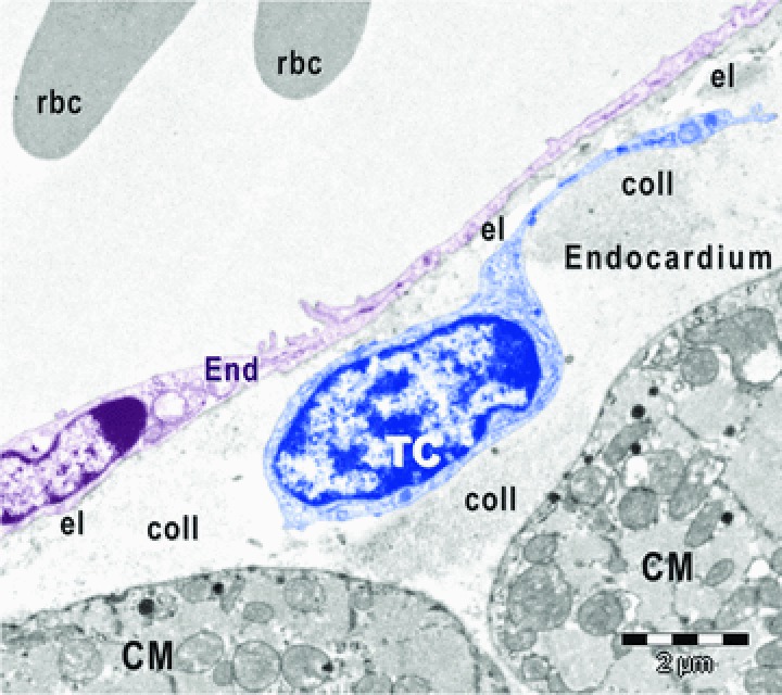
Digitally coloured electron micrograph of mouse atrial endocardium shows a telocyte (TC, blue) beneath endothelial cells (End, burgundy). CM, cardiomyocytes; rbc, red blood cells; el, elastic fibres; coll, collagen fibres.
Official websites use .gov
A
.gov website belongs to an official
government organization in the United States.
Secure .gov websites use HTTPS
A lock (
) or https:// means you've safely
connected to the .gov website. Share sensitive
information only on official, secure websites.
Digitally coloured electron micrograph of mouse atrial endocardium shows a telocyte (TC, blue) beneath endothelial cells (End, burgundy). CM, cardiomyocytes; rbc, red blood cells; el, elastic fibres; coll, collagen fibres.