
Fig 3.
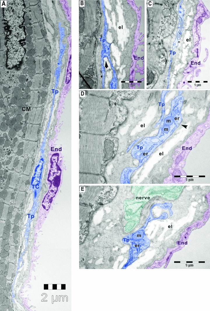
Digitally coloured electron micrographs of mouse atrial endocardium (End, burgundy). Small amount of elastin (el) is visible beneath the endothelial cells (burgundy). (A) Electron micrograph of mouse atrial endocardium shows a 30-μm-long telocyte (TC, blue) with two thin (50–500 nm) telopodes (Tp) beneath endothelial cells of endocardium. (B) Telopode with dichotomous pattern of branching (arrow). (C) Telopode with a sinuous segment having less than 50 nm thickness. (D) Dilatation of telopode hosting mitochondria (m), endoplasmic reticulum (er) and caveolae (arrowhead). (E) Telopode in close apposition with a nerve ending (n); CM, cardiomyocytes.