
Fig 4.
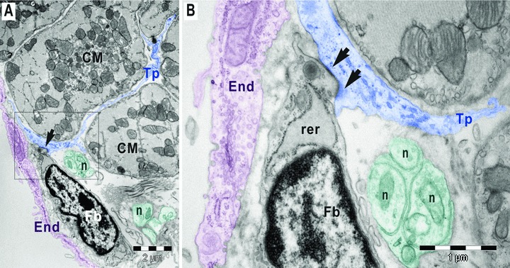
(A,B). Digitally coloured electron micrographs of mouse atrial endocardium (End, burgundy). Square marked area in A is enlarged in B. A gap junction (arrows) connects a subendocardial fibroblast (Fb) with a telopode (Tp) of a myocardial telocyte. The fibroblast has large rough endoplasmic reticulum (rer). The nerve endings (n) could be observed in between telopode and fibroblast; CM, cardiomyocytes.