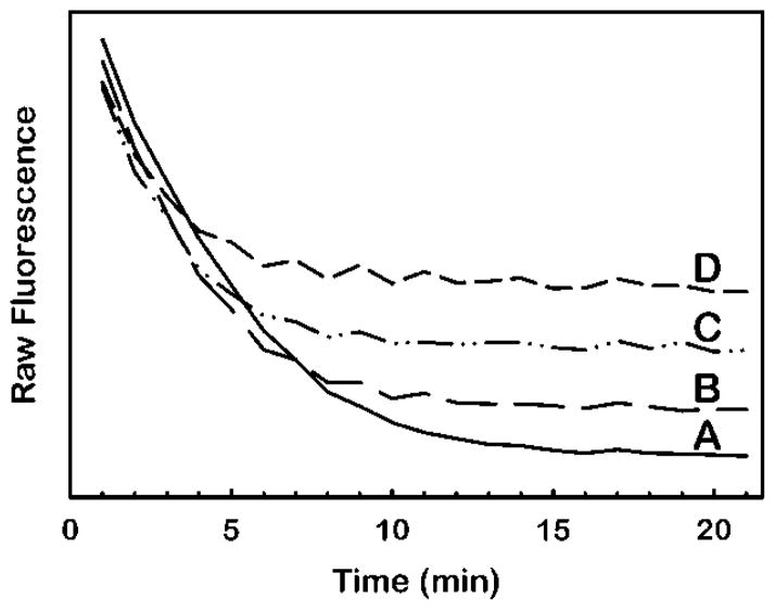
Figure 3.
Unmasking of dehydroergosteryl ester fluorescence by oxidation of excess dehydroergosterol substrate with cholesterol oxidase. For each curve, separate 100 μL volumes of HDL-mimetic substrate containing 100 μM dehydroergosterol in the assay were combined with rhLCAT to final concentrations of 0 μg/ml (A), 1 μg/ml (B), 2.4 μg/ml (C) or 4.5 μg/ml (D) and incubated for 1 hour at 37° C (Materials and Methods). Next, at 0 minutes in the figure, 25 μL of stop solution containing 5 units cholesterol oxidase per ml of 7% Triton X100 in buffer was added to each sample. The fluorescence at 325 nm excitation and 425 nm emission was monitored over time at 37° C. The higher terminal fluorescence at 20 min in (B), (C) and (D) relative to (A) is due to dehydroergosteryl ester fluorescence.

