Fig. 4.
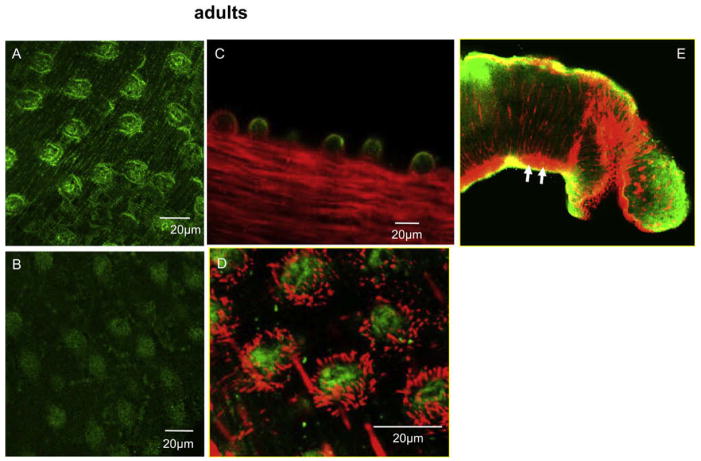
Localization of SmGPCR in adult worms. Adult male S. mansoni were probed with anti-SmGPCR IgG or a pre-adsorbed anti-SmGPCR IgG control, followed by green FITC labeled secondary antibody. Red TRITC labeled phalloidin was used to label the muscles and the tegumental spines. Animals incubated with antiserum show strong immunoreactivity in the tubercles of the dorsal tegument (A and C), whereas only background fluorescence could be detected in the negative control (B). Co-labeling with anti-SmGPCR and phalloidin produced a distinctive pattern of green immunoreactive tubercles surrounded by red phalloidin–labeled tegumental spines (D). Co-localization of SmGPCR with phalloidin was detected in the subtegumental musculature, particularly near the anterior head region (E, yellow fluorescence).

