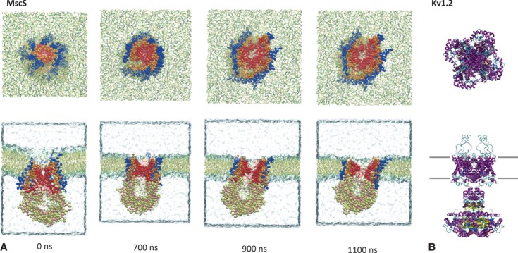
Fig. 2.

Structure of mechanosensitive ion channels. Upper panels: top views, lower panels: side views. (A) Opening of the bacterial mechanosensitive channel of small conductance (MscS, PDB accession number: 2OAU) in response to lipid bilayer stretch obtained by coarse-grained molecular dynamics simulation. MscS channel is embedded in lipid bilayers consisting of palmitoyloleoylphosphatidylcholine (POPC) and palmitoyloleoylphosphatidylethanolamine (POPE) lipids, which are shown in stick representation. The simulation box is filled with water molecules. Duration after applying a bilayer stretch is indicated in nanoseconds. Note that the channel's pore enlarges over time. (B) Three-dimensional crystal structure of mammalian Kv1.2 channel (PDB accession number: 3LUT) that putatively possesses mechanosensitivity.
