
fig 7.
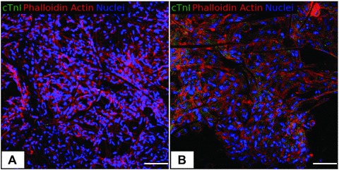
Confocal microscopy. Immunofluorescence staining for cTnI, F-actin and nuclei. (A) PLLA/Ctrl seeded with C2C12 after 48 hrs (400×). (B) PLLA/GCSF seeded with C2C12 after 48 hrs (400×). cTnI expression was found only in the PLLA/GCSF sample, with a cytoplasmatic and granular pattern of expression, that indicates a non-mature organization of the protein inside the cells. Scale bar: 100 μm.