fig 3.

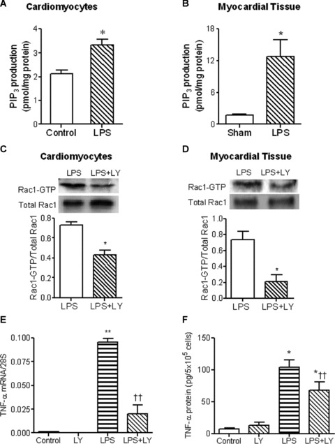
Role of PI3K in LPS-induced Rac1 activation and TNF-α expression in neonatal cardiomyocytes. Cultured WT neonatal cardiomyocytes was stimulated with LPS at 1 μg/ml for 30 min. WT mice were treated with LPS (2 mg/kg, i.p.) for 30 min. PI3K activities in cardiomyocytes (A) and myocardium (B) were determined by competitive ELISA. Rac1 activities in WT cardiomyocytes (C) and myocardium (D) stimulated with LPS for 30 min. in the presence or absence of the PI3K inhibitor LY294002 (10 μM and 7.5 mg/kg, i.p.) were determined by EZ-Detect Rac1 activation kit. WT cardiomyocytes were treated with LPS (1 μg/ml) in the presence or absence of the PI3K inhibitor LY294002 for 3 or 5 hrs. TNF-α mRNA (E) and TNF-α protein in culture medium (F) were measured by real-time RT-PCR and ELISA, respectively. Data are means ± S.E.M. from three to five independent experiments. *P < 0.05, **P < 0.01 versus control or sham; †P < 0.05, ††P < 0.01 versus LPS.
