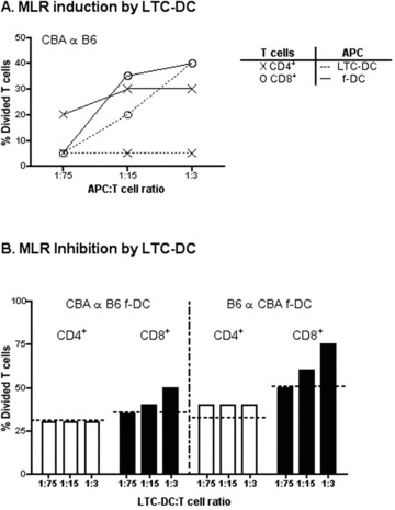
fig 3.

Ability of LTC-DC to induce and inhibit an MLR. T cells were purified from CBA/H or C57BL/6J mouse spleen and CFSE labelled. APC included freshly isolated CD11c+ spleen DC (f-DC) from CBA/H or C57BL/6J mice, or LTC-DC (B6.SJL origin). (A) Diluting numbers of APC were incubated with T cells (105) for 4 days. Cells were collected stained with antibody, and analysed flow cytometrically to determine %CD4+ or %CD8+ T cell division based on decrease in CFSE labelling. T cells cultured with no APC gave 0% division (not shown). (B) MLR co-cultures were established as in (A) using a T cell : f-DC ratio of 15:1. Varying numbers of LTC-DC were added into co-cultures to test their ability to inhibit the MLR reaction. Percentage T-cell division was calculated as in (A), and the level achieved in the absence of added LTC-DC is shown as a dashed line.
