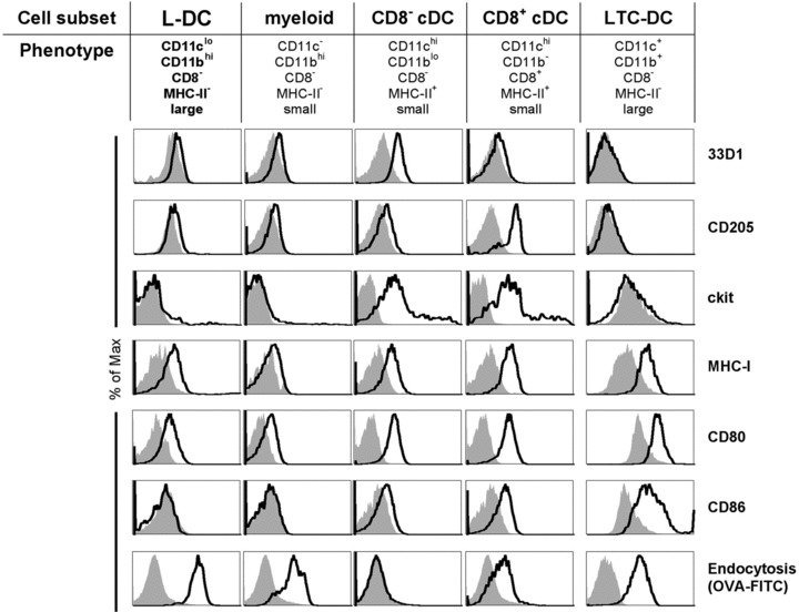
fig 5.

Phenotypic characterization of L-DC. APC subsets including L-DC were identified flow cytometrically according to the gating procedure in Figure 4 using CD11c, CD11b, CD8α, MHC-II and FSC (size) and further assessed for marker expression. In vitro-derived LTC-DC were stained for comparison. Prior to flow cytometry, cells were incubated with PI (1 μg/ml) for gating of live (PI−) cells. In order to measure the in situ capacity of cells for endocytosis, mice were given FITC-OVA (3 mg) i.v. 24 hrs prior to isolation of spleen cells for antibody staining and analysis. Endocytosis was assessed for each gated subset in relation to a non-endocytic control spleen lymphocyte population (filled histogram). For LTC-DC, endocytosis was measured after in vitro treatment with OVA-FITC for 45 min. at 37°C or at 4°C as a control.
