fig 3.

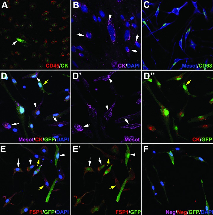
Antigen immunolocalization in adherent cells obtained from peritoneal lavages 48 hrs after injury of the peritoneal wall. (A–C) show cells obtained from normal BALB/c mice whereas (D)–(F) show cells from mice reconstituted with GFP-expressing bone marrow. (A) Virtually all the CD45+ cells show a perinuclear dot-like pattern of cytokeratin immunoreactivity. Note a strongly CD45–/cytokeratin+ immunoreactive cell, probably a delaminated mesothelial cell (arrow). (B) The perinuclear dot-like pattern of cytokeratin immunoreactivity is also present in mesothelial cells migrating from omentum explants, which were used as positive controls (arrows). Other cells still show an extended cytokeratin cytoskeleton (arrowhead). (C) Mesothelin immunoreactivity colocalized with CD68. Note the cytoplasmic and perinuclear CD68 localization. (D) Triple localization of mesothelin, cytokeratin and GFP. This immunostaining revealed different phenotypes, including triple positive cells (arrowheads), mesothelin+/cytokeratin+, GFP– cells which probably are delaminated mesothelial cells (white arrows) and GFP+, mesothelin–/cytokeratin– cells (yellow arrow). (E) The fibroblast marker FSP1 was expressed in many GFP+ cells (white arrows), as well as in GFP– cells (yellow arrows). Other GFP+ cells were negative for FSP1 (arrowhead). (F) Negative control incubated with isotype primary antibodies and with the same secondary antibodies as used in the rest of the figures.
