
fig 5.
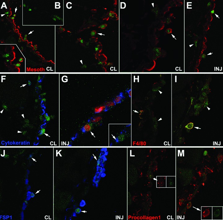
Colocalization of GFP and mesothelin, cytokeratin, F4/80, FSP1 and procollagen-1 in the injured (INJ) or contralateral (CL) peritoneal wall from mice reconstituted with GFP-expressing bone marrow, 48 hrs after surgery. (A–D) Colocalization of GFP and mesothelin in the contralateral areas. Some double-labelled cells are apparently detaching from the mesothelial surface (arrows). GFP+ cells within the tissue are mesothelin– as shown in (B). GFP+/mesothelin– cells are also abundant in sub-mesothelial areas (arrowheads). (E) Double labelled cells can also be seen in the regenerating mesothelium of the injured surface (arrows). GFP+/mesothelin– cells are also present but they are less abundant than in contralateral areas (arrowhead). (F), (G) Colocalization of GFP and cytokeratin can be observed in the contralateral areas (arrows in F) but it is apparently scarcer in the injured ones (G). GFP+/cytokeratin– cells are shown in the contralateral side (arrowheads in F). (H), (I) The macrophage marker F4/80 is present in a few cells from both areas (arrows). However, most of the apparently adhered GFP+ cells in the injured area do not express this macrophage marker (arrowheads). (J), (K) Colocalization of GFP with the fibroblastic marker FSP1 (arrows). (L), (M) Colocalization of GFP with procollagen-1 (arrows).