
fig 6.
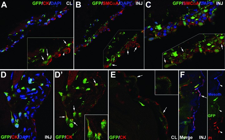
Colocalization of GFP, mesothelial and fibroblastic markers in the contralateral (CL) and injured (INJ) peritoneal wall from mice reconstituted with GFP-expressing bone marrow, 1 week (A–C) and 1 month (D–F) after surgery. (A) Colocalization of cytokeratin and GFP in the injured area 1 week after surgery. Some GFP+ cells show an extended cytokeratin cytoskeleton (arrowhead) whereas others show the perinuclear dot-like cytokeratin pattern (arrows). (B), (C) Colocalization of smooth muscle cell α-actin with GFP in the injured area 1 week after surgery. GFP+ cells are very abundant and form several cell layers covering all the damaged area. Most of these cells express smooth muscle cell α-actin. (D–F) Colocalization of mesothelial markers and GFP in the injured and contralateral areas 1 month after surgery. The mesothelium is completely regenerated. GFP+ cells are present in the sub-mesothelial area, and some of them show a perinuclear cytokeratin dot (arrows in D’). It is still possible to find a few GFP+ cells integrated in the mesothelial lining of both, injured and contralateral areas, expressing cytokeratin (arrows in E) and mesothelin (arrows in F).