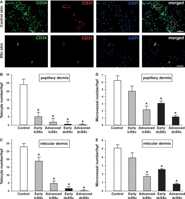
Fig. 5.

Quantitative analysis of telocytes and microvessels in skin sections from controls and systemic sclerosis (SSc) patients double-immunolabelled for CD34 (green) and CD31 (red) and counterstained with DAPI (blue) for nuclei. (A) Representative photomicrographs from control and clinically affected SSc skin samples are shown. Scale bar = 50 μm. CD34-positive/CD31-negative spindle-shaped cells (telocytes) and CD34/CD31-double-positive microvessels were counted in 10 randomly chosen microscopic high-power fields (hpf; 40× original magnification) of the papillary dermis (B and D) and 10 hpf of the reticular dermis (C and E) per sample. Only the cells with well defined nuclei were counted. Data are represented as mean ± SD. *P < 0.05 vs control (by Student's t-test). Limited cutaneous SSc: lcSSc; diffuse cutaneous SSc: dcSSc.
