
Fig. 9.
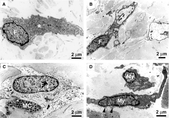
Diffuse cutaneous systemic sclerosis (dcSSc) skin, transmission electron microscopy. (A) Early dcSSc. An activated fibroblast displays a particularly extended cytoplasm containing an abundant rough endoplasmic reticulum and a large Golgi apparatus (asterisk). (B and C) Advanced dcSSc. Some fibroblasts embedded in the fibrotic extracellular matrix show ultrastructural features suggestive of a functional exhaustion, such as extremely dilated endoplasmic reticulum cisternae (B) or a clear and empty cytoplasm (C). Note a mast cell in close contact with the fibroblast (C). (D) Advanced dcSSc. Myofibroblasts show a large body with short and thick processes and are rich in cisternae of rough endoplasmic reticulum, mitochondria and myofilaments. Subplasmalemmal focal densities are evident (arrowheads). F: fibroblast; MC: mast cell; My: myofibroblast. Scale bars are indicated in each panel.