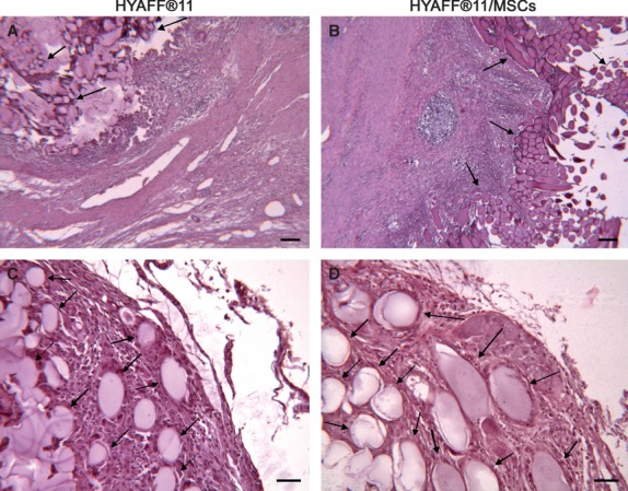
Fig. 3.

Morphology of HYAFF®11 fibres transplanted in the infarct area. The contour of the HYAFF®11 fibres from both HYAFF®11 and HYAFF®11/MSCs groups is oval or lengthened, according to the direction of the mesh. These samples were obtained 4 weeks after transplantation and were stained with haematoxylin and eosin. Scale bars: (a and b) 100 μm; (c and d) 50 μm.
