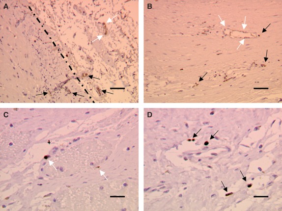
Fig. 5.

Localization of BrdU-labelled MSCs. In the myocardial preparations, the dark brown elements identified the nuclei of MSCs pre-loaded with BrdU before implantation. (a) BrdU-labelled MSCs were mainly present in the infarct region and in the border zone (dotted line), whereas few of them moved to the healthy remote regions of the heart (data not shown). (b–d) Many BrdU+MSCs were localized at the level of vessels structures, just near them (black arrows), or inside the vessel wall (white arrows). Scale bars: (a and b) 25 μm, (c and d) 10 μm.
