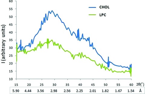
Fig 3.

Wide-angle X-ray diffraction pattern of interacting t- and v-SNARE-reconstituted lipid vesicles, with the vesicle membrane containing either cholesterol (CHOL) or lysophosphatidylcholine (LPC). Diffraction profiles of a mixture of 50 nm in diameter t-SNARE and v-SNARE reconstituted PC:PS vesicles either containing cholesterol CHOL or LPC. Note as a consequence of clustering of LPC-containing vesicles, the diffraction is significantly lower (green peak) compared to CHOL-containing vesicles (blue peak). Interestingly, the distance between interacting t-SNARE and v-SNARE vesicles is closer between the CHOL populations (3.05 Å) compared to LPC (3.33 Å).
