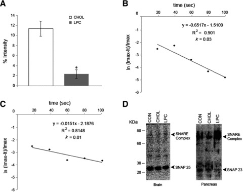
Fig 4.

SNARE complexes in the presence of LPC fail to disassemble. (A) Real-time dynamic light scattering (DLS) profiles on cholesterol-associated (CHOL) and LPC-associated t-/v-SNARE liposomes in presence of NSF–ATP. There is no appreciable dissociation of the LPC vesicles, in contrast to a rapid ATP-dependent dissociation of CHOL vesicles (P < 0.001). (B) Note the dissociation of cholesterol-associated t-/v-SNARE vesicles occurs with rate constant k = 0.03 sec.−1, and (C) a slow dissociation in LPC vesicles (k = 0.01 sec.−1). (D) Following KCl stimulation, isolated brain slices pre-incubated in CHOL, LPC or vehicle (CON) are solubilized in buffer containing ATP–EDTA, and 10 mg of protein resolved by SDS-PAGE, followed by immunoblot analysis using SNAP-25-specific antibody, negligible disassembly of the t-/v-SNARE complex is demonstrated in brain tissue pre-incubated in LPC, as opposed to the control (CON) or CHOL. Similarly, exocrine pancreas pre-incubated in LPC, demonstrate reduced disassembly of the t-/v- SNARE complex following stimulation of secretion using 1 mM carbamylcholine.
