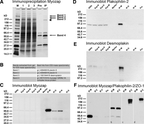
Fig 3.

Results of immunoprecipitation experiments using myozap mAb (clone 517.67) and mouse cardiomyocyte HL-1 cell lysates. (A) SDS-PAGE of polypeptides of fractions obtained in immunoprecipitation experiments as seen after silver staining: lane M: broad range Mr reference molecules; lane 1: initial RIPA buffer lysate; lane 2: supernatant of the ‘preclearing step’; lane 3: supernatant after myozap immunoprecipitation; lane ‘Pre’: polypeptides of the ‘preclearing step’ absorbed to dynabeads; lane ‘IP’: lysate of myozap immunoprecipitate. Bands 1–4 denoted by arrows have been used for electron spray ionization (ESI) mass spectometry. Band 4, arrow denotes the band containing both myozap as well as residual desmin and IgG heavy chain. Arrowhead, immunoglobulin (IgM heavy chain residual contamination). (B) Table of results of ESI mass spectometry of the extracted bands 1–4 (see arrows in A). Note that, together with myozap (band 4), three other proteins are detected in the immunoprecipitates: desmoplakin (in band 3), plectin (in bands 1 and 2) and the intermediate filament protein desmin (in band 4). (C) Immunoblot reactions with myozap mAb: lane ‘RIPA’: initial RIPA buffer lysate; lane ‘Sup-E’: supernatant of immunoprecipitation using E-cadherin antibodies; lane ‘Sup-N’: supernatant of N-cadherin immunoprecipitation; lane ‘Sup-D’: supernatant of desmoglein-2 (Dsg-2) immunoprecipitation; lane ‘Sup-M’: supernatant of myozap immunoprecipitation; lane ‘Marker’: broad range protein Mr markers (New England Biolabs, Frankfurt, Germany); Mr values are indicated on the left margin; lane ‘IP-M’: myozap immunoprecipitate; lane ‘IP-D’: desmoglein-2 immunoprecipitate; lane ‘IP-N’: N-cadherin immunoprecipitate; lane ‘IP-E’: E-cadherin immunoprecipitate (negative control). (D, E) Fractions as in B, showing immunoblot reactions with antibodies to plakophilin-2 (D) and desmoplakin (E). Lane designations as in C. Note that under these conditions immunocomplexes of myozap with desmoplakin and plakophilin-2 are identified. (F) Specific parallel immunoprecipitations using antibodies to protein myozap (IP-M; F, a), protein ZO-1 (IP-Z; F, a), VE-cadherin (IP-V; F, a), in comparison with the ‘pre-clearing’ pellet (‘Pre’; see also above) as negative control. For comparison, immunoprecipitates obtained with antibodies to plakophilin-2 (b) and to protein ZO-1 using RIPA buffer (c) or Triton-X100-containing buffer (d) are shown. The specific immunoblot reactions with antibodies to myozap (a), plakophilin-2 (b) and protein ZO-1 (c, d) are shown.
