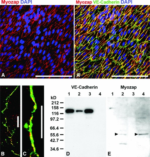
Fig 6.

Bovine aortic endothelium in a fresh detached cell layer preparation showing the reaction of protein myozap in the zonulae adhaerentes of a bovine aorta. (A) Immunolocalization of myozap (red) in the detached endothelium. (A’) Same region as in (A) but in an optical channel showing both VE-cadherin (green) and myozap (red) reactions with far-reaching colocalization (yellow merge colour). (B, C) Laser-scanning double-label immunofluorescence microscopy of cultured human endothelial HUVEC cells, showing the partial association of myozap (red) with other adherens junction proteins such as (both in green) β-catenin (B) or VE-cadherin (C). (D, E) Results of fractionation experiments of HUVEC cell lysate proteins after SDS-PAGE of polypeptides and immunoblotting with antibodies to VE-cadherin (D) and myozap (E) are shown. Presented are proteins of the supernatants (lanes 1 and 3) and the remaining pellet fractions (lanes 2 and 4) after lysis using Triton-X100 (lanes 1 and 2) or RIPA (lanes 3 and 4) buffer, showing the relative recoveries of VE-cadherin (D) in comparison with protein myozap (E) which is predominantly detectable in the pellet fractions of the detergent-containing protein lysates (arrowheads). The masses of the molecular size markers (kD) are presented on the left margin of D. Bars: 100 μm (A, A’); 20 μm (B, C).
