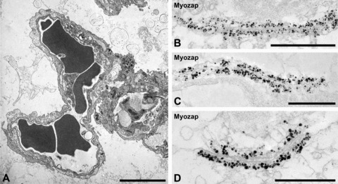
Fig 7.

(A) Transmission electron micrographs of sections through mouse lung alveoli. Note the close neighbourhood of capillary endothelial (left part) and respiratory alveolar epithelial structures (right part) structures (erythrocytes appear as dark cells in the lumen of microcapillaries). (B–D) Single-label immunoelectron microscopy showing the specific ultrastructural localization of protein myozap in the zonulae adhaerentes connecting endothelial cells. Note that the cytoplasmic plaques of the endothelial AJ system are entirely and strongly decorated by myozap-antibodies coupled to colloidal gold particles (with silver enhancement), whereas such labelling is absent from the membrane contact region and the intercellular connection structures (D). Bars: 5 μm (A), 1 μm (B), 0.5 μm (C and D).
