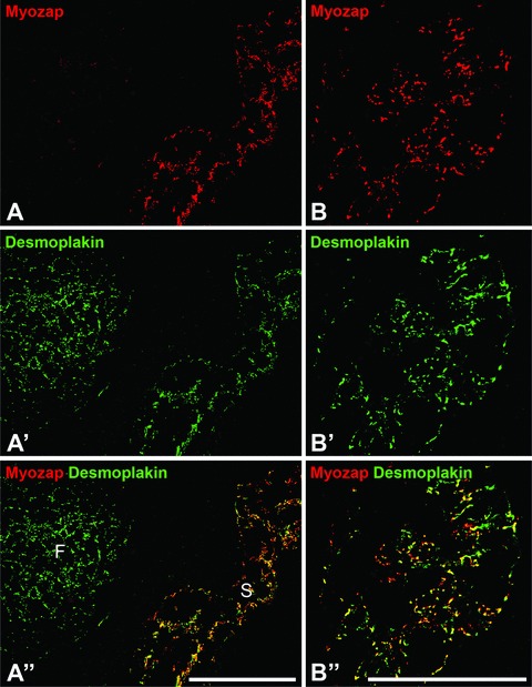
Fig 8.

Double label immunofluorescence microscopy of protein myozap in junctions connecting the endothelial cells of the lymphatic vessel system, here on cryostat sections through bovine (A) and human (B) lymph nodes. Note that the complexus adhaerens junctions connecting the endothelial cells of the virgultar arrangements in the lymph node sinus regions are positive for myozap (red in A and B) and desmoplakin (green in A’ and B’) resulting in yellow merge labelling (A’’ and B’’). In contrast, the desmosomes of the follicular dendritic reticulum (F) are strongly positive for desmoplakin only (green in A’ and A’’). Bars: 100 μm.
