Fig 1.

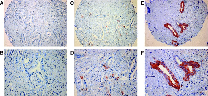
Immunohistochemical staining of HSP27 in pancreatic cancer. Representative microscopic pictures of pancreatic ductal adenocarcinomas considered negative (A and B), weakly positive (C and D) or strongly positive (E and F).

Immunohistochemical staining of HSP27 in pancreatic cancer. Representative microscopic pictures of pancreatic ductal adenocarcinomas considered negative (A and B), weakly positive (C and D) or strongly positive (E and F).