Fig 5.

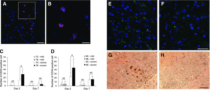
Representative pictures of CD34+ cells in ventral grey matter (A) and enlargement of boxed area (B). Blue: nuclei and red: CD34. (A) Scale bar = 25 μm; (B) Scale bar = 10 μm. The number of CD34+ cells in the lumbar spinal cord after 7-min. (C) and 9-min. SCI (D). CD34+ cells were more abundantly recruited in the spinal cord in groups 7E-severe and 9E-severe than those in groups 7C-severe and 9C-severe at day 2 (C and D, *P < 0.05 with Bonferroni correction). CD34+ cells were not apparent in groups 7C-mild, 7E-mild, 9C-mild and 9E-mild. Representative pictures of immunofluorescence for BDNF in ventral grey matter from groups 9E-severe (E) and 9C-severe (F) at day 7. Blue: nuclei and green: BDNF. BDNF expression in group 9E-severe (E) was more evident than that in group 9C-severe (F). Scale bar = 25 μm. Representative pictures of VEGF expression in the left ventrolateral white matter of the spinal cord from groups 9E-severe (G) and 9C-severe (H) at day 7. VEGF was detectable in the white matter of group 9E-severe and vacuolization in white matter was limited (G). In contrast, no VEGF expression and advanced vacuolization was apparent in the white matter of group 9C-severe (H). Scale bar = 50 μm.
