Fig 2.

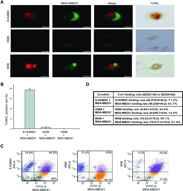
MDA-MB231 apoptosis following binding or formation of cell-in-cell structure with selected HUMSC. (A) Confocal microscopy demonstrated the cell-in-cell structure of selected HUMSC (red) internalized within MDA-MB231 (green) after co-culture. Simultaneous presentation of strong red fluorescence came from HUMSC within the region of green fluorescence delineated by MDA-MB231 suggested that some substance from HUMSC had been intermixed within MDA-MB231, and the MDA-MB231 was stained positively by TUNEL (top panels). Binding of HS68 (red) with MDA-MB231 (green) was rare, and no cells were TUNEL-positive (middle panels). The cell-in-cell structure of WI38 (red) internalized within MDA-MB231 (green) was stained negatively by TUNEL (bottom panels). Scale bars: 10 μm. (B) The percentage of TUNEL-positive cells after co-culture for 3 days was 17.66 ± 0.58% of co-cultured MDA-MB231 with selected HUMSC, but only 0.61 ± 0.31% of co-cultured MDA-MB231 with WI38 and 0 ± 0% of co-cultured MDA-MB231 with HS68 was TUNEL-positive. Data are means ± SEM. (C) Using flow cytometry, the number of cells with simply red (Q1), simply green (Q4) and dual (red + green) fluorescence (Q2) was measured. The percentage of cells with dual fluorescence was 49.2%, 9.9% and 79.2% after co-culture of MDA-MB231 with selected HUMSC, HS68 and WI38, respectively. (D) The binding rate of each cell population.
