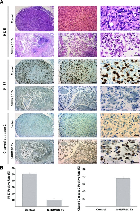
Fig 5.

Histopathologic evidence of the suppression of breast cancer tumourigenesis by selected HUMSCs. (A) On haematoxylin and eosin staining, the tumour cells in breast cancer control group (n = 7) were dense and tight, but the tumour cells in HUMSC treatment group (n = 7) were sparse and loose, and condensation of nuclei was observed. On immunohistochemical staining of KI-67, lots of positive cells were noted within the tumour nodule in breast cancer control group (n = 7), but very few positive cells were found in HUMSC treatment group (n = 7). On immunohistochemical staining of cleaved-caspase-3, very few positive cells were found in breast cancer control group (n = 7), but lots of positive cells were noted within the tumour nodule in HUMSC treatment group (n = 7). Scale bars: 20 μm (right) or 100 μm (all others). (B) The KI-67 positive staining rate under high power field of the tumour nodule was 51.21 ± 1.72% in breast cancer control group but was only 10.47 ± 1.61% in HUMSC treatment group. The cleaved-caspase-3 positive staining rate under high power field of the tumour nodule was 0 ± 0% in breast cancer control group but was 75.04 ± 4.65% in HUMSC treatment group.
