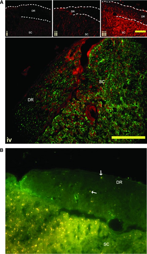
Fig 5.

(A) BDNF immunoreactivity in the dorsal roots. Representative sections of dorsal root entry zones from the lumbar spinal cord of Lewis rats induced to a state of EAE. A marked increase in BDNF immunoreactivity (red) is seen in the dorsal roots of active EAE compared to active control animals and NC animals. Panel i—Naïve animals have no BDNF immunoreactivity in the dorsal roots (DR). Panel ii—Active control animals have noticeable BDNF immunoreactivity in the satellite cells of the DR, and in the grey matter of the dorsal horn. Panel iii—Active EAE animals appear to have increased levels of BDNF immunoreactivity in the satellite cells of the DR, as identified as small brightly stained cell surrounding the axons of the root. In addition, there is a marked increase in punctuate staining in the length of the roots. Further, the grey matter of the dorsal horn has a markedly increased immunoreactivity compared to the active control animals. DR = Dorsal root; SC = spinal cord. Bar = 10 μm. Panel iv—BDNF expression (red) is associated with neurons (labelled with Neuron-specific class III beta-tubulin (Tuj1) – green) in both the DR, and the dorsal horn. Bar = 10 μm (B) Anterograde transport of BDNF along the dorsal root is mediated by kinesin. Double immunolabelling of frozen sections of EAE spinal cord shows BDNF (red) co-localized to the motor protein kinesin (green) (arrow). Kinesin is distributed throughout the dorsal root (DR) and spinal cord (SC). BDNF immunoreactivity is markedly higher in the dorsal root entry zone than the root. Co-labelling shows as yellow. Bar = 10 μm.
