Fig 2.

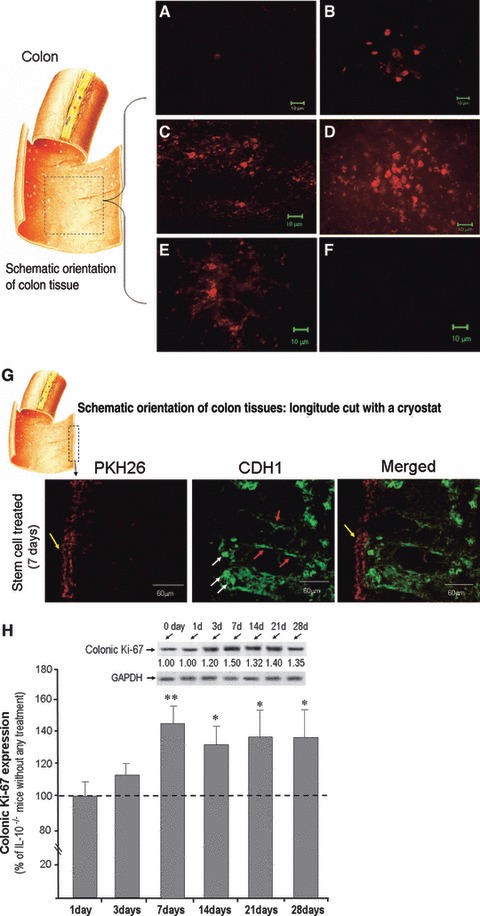
Co-localization of CSCs in inflamed colon: An additional group of mice was used for detecting stem cell location. Locally transplanted CSCs and CECs were labelled using PKH26 red fluorescent to detect stem cell migration to the inflamed colon in IL-10−/− mice. A piece of mouse colon (∼1 cm) was removed and the colon was opened (see left panel of colon picture), and then mounted flat with the mucosal surface facing up. The fluorescent signals from CSCs were predominantly detected in the mucosa of the inflamed colon. (A) 1 day after stem cells local infusion; (B) 3 days after stem cells local infusion; (C) 7 days after stem cells local infusion; (D) 14 days after stem cells local infusion; (E) 28 days after stem cells local infusion. No fluorescent signal was found after 14 days local infusion of CECs (F). The stem cell treated mouse colon with PKH26 label was also selected which was longitude cut with a cryostat, and then double labelled with E-cadherin (CDH1). As shown in (G), following a longitude cutting a colon tissue, the stem cell with PKH26 signal was presented in the surface of mucosa (yellow arrows) 3 days after stem cell transplantation, and CDH1 was showed in intracellular compartments (white arrows), and lateral membrane of crypts (red arrows). (H) shows Western blots indicated Ki-67 expression in CSCs treated mice (H, upper panel) compared to IL-10−/− mice without any treatment. There are six time points (1, 3, 7, 14, 21 and 28 days following CSCs infusion, each time point representative of 3 mice, and plus 3 IL-10−/− mice without any treatment as indicated). There was a significant up-regulated of the Ki67 expression was detected from 7 days following CSCs infusion to 4 weeks (H, lower panel).
