
Fig 1.
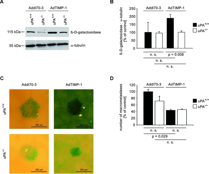
META/Bomnu/nu mice of variable uPA genotype (uPA+/+ and uPA−/−, respectively) were transduced by either AdTIMP-1 or Addl70–3 adenoviruses and 3 days later challenged with lacZ-tagged L-CI.5s cells. Killing and liver removal were performed another 6 days later. (A) Representative Western blot of β-D-galactosidase, the protein encoded by the tumour cell tag lacZ, in liver protein. (B) Densitometry of all performed western blots revealed increased total tumour cell mass in livers with elevated TIMP-1. Loss of host uPA diminished this augmented metastatic burden. Columns: Mean intensities of the β-D-galactosidase bands versusα-tubulin band intensities. The mean of the reference group Addl70–3/uPA+/+ was set as 100%. Bars: S.E. n= 5 mice. Addl70–3/uPA+/+: 100.0% 6 27.7%; Addl70–3/uPA−/−: 95.0%± 4.3%; AdTIMP-1/uPA+/+: 188.1%± 27.7%; AdTIMP-1/uPA−/−: 100.5%± 3.7%. (C) Close-up pictures of surfaces of representative X-Gal-stained metastasis-bearing livers showed increased scattering of tumour cells (indigo-blue signal) in livers with elevated TIMP-1 levels. Loss of host uPA led to a subtotal reduction of this micrometastatic spread. (D) Quantification of macrometastases in X-Gal-stained metastasis-bearing livers revealed a reduction of multicellular foci in livers with elevated TIMP-1 levels. Columns: Mean number of macrometastases. The mean of the reference group Addl70–3/L-CI.5s was set as 100%. Bars: S.E. Addl70–3/uPA+/+: 100.0%± 7.5%, n= 3 mice; Addl70–3/uPA−/−: 71.9%± 14.0%, n= 5 mice; AdTIMP-1/uPA+/+: 44.3%± 2.7%, n= 4 mice; AdTIMP-1/uPA−/−: 46.1%± 1.8%, n= 4 mice.