
Fig 2.
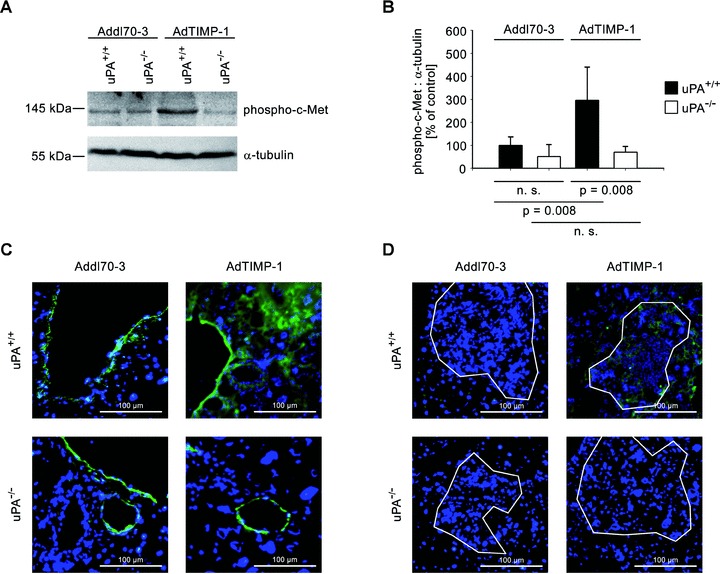
(A) Representative western blot of phosphorylated c-Met in liver protein of META/Bomnu/nu mice (uPA+/+ and uPA−/−, respectively), transduced by either AdTIMP-1 or Addl70–3 adenoviruses. (B) Densitometry of all performed Western blots revealed increased c-Met activation in livers with elevated TIMP-1. Loss of host uPA diminished this induction of HGF signalling. Columns: Mean intensities of the phospho-c-Met bands versus α-tubulin band intensities. The mean of the reference group Addl70–3/uPA+/+ was set as 100%. Bars: S.E. n= 5 mice. Addl70–3/uPA+/+: 100.0%± 16.1%; Addl70–3/uPA−/−: 51.5%± 22.9%; AdTIMP-1/uPA+/+: 296.7%± 63.9%; AdTIMP-1/uPA−/−: 69.9%± 11.2%. (C and D) Representative immunofluorescence analysis of phospho-c-Met (green signal) on cryo-sections (5 μm) of metastasis-bearing bearing livers. Counterstaining was done with DAPI (blue signal). Approximate boundaries of macrometastases were delineated in white. (C) In both Addl70–3- and AdTIMP-1-transduced livers strong staining of phospho-c-Met was found around veins. In livers with elevated TIMP-1, additional c-Met activation in parenchyma was detected. Lack of host uPA did only affect the parenchymatous localization of HGF signalling. (D) Within and around macrometastases, only in livers of uPA wild-type animals with elevated TIMP-1 levels HGF signalling was induced.