
Fig 6.
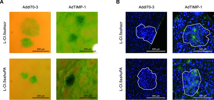
(A) Close-up pictures of surfaces of representative X-Gal-stained metastasis-bearing livers of CD1nu/nu mice, transduced by either AdTIMP-1 or Addl70–3 adenoviruses, showed increased scattering of tumour cells (indigo-blue signal) in livers with elevated TIMP-1 levels. Lack of tumour cell uPA did not reduce micrometastatic spread. (B) Representative immunofluorescence analysis of phospho-c-Met (green signal) on cryo-sections (5 μm) of metastasis-bearing livers. Counterstaining was done with DAPI (blue signal). Approximate boundaries of macrometastases were delineated in white. In both Addl70–3- and AdTIMP-1-transduced livers strong staining for phospho-c-Met was found within and around macrometastases. Lack of tumour cell expression of uPA did not impair this induction of HGF signalling.